Fiberoptic Bronchoscopy: A Patient's Guide to the Procedure
Key points
- Flexibility: A long, flexible tube constructed from woven metal braiding and polymer sheathing that can reach deep into lung segments without traumatizing the delicate bronchial walls. The tip can bend up to 210 degrees, allowing navigation through the sharp anatomical turns of the tracheobronchial tree.
- Vision: A video chip or fiber-optic bundles transmit clear, high-definition images to an external monitor. Advanced models feature narrow-band imaging (NBI) and autofluorescence technology to highlight abnormal vascular patterns or precancerous tissue that may be invisible under standard white light.
- Size: A small diameter (typically 4 to 6 mm, or roughly 5 mm) allows passage through the nose and into smaller bronchi while maintaining adequate suction and instrument passage capabilities. Thinner pediatric or ultrathin scopes (around 3 mm) exist for highly stenotic airways.
- Working Channel: A dedicated lumen (usually 1.8 to 3.0 mm in diameter) allows for the use of biopsy tools, cytology brushes, aspiration needles, balloon catheters for dilation, or suction devices to clear secretions. This versatility enables multiple interventions during a single session.
- Control: A handle with up/down and left/right deflection dials, alongside angulation locks and suction ports, allows the doctor to steer the tip with millimeter precision inside the lung while maintaining continuous visualization.
Fiberoptic bronchoscopy—often called just bronchoscopy—is a common, minimally invasive procedure that allows doctors to look inside your airways and lungs. If you or a loved one has been advised to undergo this procedure, it's natural to have questions. Respiratory conditions can be complex, and modern pulmonology relies heavily on direct visualization to differentiate between infections, inflammatory diseases, structural abnormalities, and malignancies. This guide explains what a fiberoptic bronchoscopy is, why it's done, how to prepare, and what to expect during and after. By understanding the clinical rationale, procedural steps, and recovery timeline, patients can approach their appointment with confidence and clarity.
What is Fiberoptic Bronchoscopy?
Fiberoptic bronchoscopy is a medical procedure where a doctor uses a thin, flexible tube with a light and camera (a bronchoscope) to examine your respiratory tract, including the windpipe (trachea) and the branching airways (bronchial tubes) of the lungs. The term "fiberoptic" refers to the flexible technology that transmits light and images, allowing the scope to navigate the natural curves of your airways. It is also known as a flexible bronchoscopy. While early versions relied strictly on glass fiber bundles to relay images, modern iterations often incorporate a high-resolution digital chip at the distal tip, though the traditional fiberoptic terminology remains widely used in clinical practice and patient education.
The bronchoscope, about the diameter of a pencil, is gently inserted through the nose or mouth and guided down the throat into the lungs. The tiny camera at its tip sends real-time video to a monitor, giving the doctor a clear view of the airway lining. The scope also has a channel for passing tiny instruments to take a tissue sample (biopsy), collect cells with a brush, or remove mucus. This working channel is a critical feature, transforming the device from a purely diagnostic viewer into an active therapeutic tool. Physicians can introduce saline, forceps, cryoprobes, balloons, and even stents through the same narrow passage, minimizing the need for open surgery.
"Fiberoptic bronchoscopy has revolutionized how we diagnose lung diseases, offering a real-time view inside the airways with minimal invasion. It allows us to gather crucial information and even perform treatments, all while keeping the patient comfortable." — A pulmonology specialist.
This procedure is typically performed by a pulmonologist (lung doctor) or a thoracic surgeon in a hospital endoscopy suite, an outpatient bronchoscopy center, or sometimes an intensive care unit. It is usually conducted under light sedation, though the depth of sedation can be tailored to patient needs, procedural complexity, and clinical setting. The technique has a high diagnostic yield and remains the gold standard for evaluating endobronchial lesions, unexplained hemoptysis, and diffuse parenchymal lung diseases.
Key Features of the Bronchoscope:
- Flexibility: A long, flexible tube constructed from woven metal braiding and polymer sheathing that can reach deep into lung segments without traumatizing the delicate bronchial walls. The tip can bend up to 210 degrees, allowing navigation through the sharp anatomical turns of the tracheobronchial tree.
- Vision: A video chip or fiber-optic bundles transmit clear, high-definition images to an external monitor. Advanced models feature narrow-band imaging (NBI) and autofluorescence technology to highlight abnormal vascular patterns or precancerous tissue that may be invisible under standard white light.
- Size: A small diameter (typically 4 to 6 mm, or roughly 5 mm) allows passage through the nose and into smaller bronchi while maintaining adequate suction and instrument passage capabilities. Thinner pediatric or ultrathin scopes (around 3 mm) exist for highly stenotic airways.
- Working Channel: A dedicated lumen (usually 1.8 to 3.0 mm in diameter) allows for the use of biopsy tools, cytology brushes, aspiration needles, balloon catheters for dilation, or suction devices to clear secretions. This versatility enables multiple interventions during a single session.
- Control: A handle with up/down and left/right deflection dials, alongside angulation locks and suction ports, allows the doctor to steer the tip with millimeter precision inside the lung while maintaining continuous visualization.
In essence, bronchoscopy lets doctors explore the lungs from the inside, enabling them to see what’s happening and intervene if needed. By combining direct visual inspection with targeted sampling, it bridges the gap between non-invasive imaging and invasive surgical thoracotomy, offering a balanced approach to respiratory diagnostics and therapy.
Why is a Bronchoscopy Done?
A bronchoscopy may be recommended for diagnosis, investigation, or treatment of various lung conditions. When imaging studies like chest X-rays or computed tomography (CT) scans reveal abnormalities that cannot be definitively characterized non-invasively, bronchoscopy provides the necessary tissue and visual confirmation. The following are the primary clinical indications, expanded for clarity:
- Persistent Cough or Wheezing: To find the cause of a chronic cough not explained by other conditions like asthma, GERD, or post-nasal drip. The procedure can identify endobronchial lesions, tracheobronchomalacia (airway collapse), or localized inflammation that standard pulmonary function tests cannot detect.
- Coughing up Blood (Hemoptysis): To locate the source of bleeding within the airways. Bronchoscopy can identify ulcerations, tumors, bronchitis, or vascular malformations causing hemorrhage. It also allows for immediate intervention, such as cold saline lavage, topical vasoconstrictors, or balloon tamponade to control active bleeding.
- Abnormal Chest X-Ray or CT Scan: To investigate a visible spot, nodule, or mass by viewing it directly and obtaining a biopsy to check for conditions like lung cancer. Transbronchial biopsies or endobronchial forceps samples provide histopathological confirmation essential for staging and treatment planning.
- Suspected Lung Infections: To collect samples (mucus or fluid) to identify specific pathogens like tuberculosis or fungi, especially in recurrent pneumonia, immunocompromised patients, or ventilator-associated pneumonia. Microbiological cultures, PCR assays, and acid-fast bacilli smears from bronchial washings significantly improve pathogen identification compared to routine sputum samples.
- Airway Blockage: To find and sometimes remove an obstruction, such as an inhaled foreign object or a tumor. In cases of benign or malignant central airway obstruction, bronchoscopic techniques like electrocautery, argon plasma coagulation, cryoextraction, or mechanical debulking can rapidly restore airflow and relieve respiratory distress.
- Interstitial Lung Disease (ILD): A procedure called bronchoalveolar lavage (BAL), where a small amount of saline is washed into the lung and suctioned back, can help diagnose conditions like sarcoidosis, hypersensitivity pneumonitis, or pulmonary alveolar proteinosis. Cellular differential analysis of the recovered fluid reveals characteristic immune cell patterns that guide targeted therapy.
- Lung Cancer Staging: A specialized technique called endobronchial ultrasound (EBUS) combines bronchoscopy with ultrasound to sample lymph nodes in the chest. EBUS-guided transbronchial needle aspiration (TBNA) is now standard for minimally invasive mediastinal staging, reducing the need for surgical mediastinoscopy while maintaining high diagnostic accuracy.
- Mucus Plugs: To suction out thick mucus that is causing a part of the lung to collapse (atelectasis), particularly in hospitalized patients, those with cystic fibrosis, or individuals with impaired cough reflexes due to neurological conditions. Rapid clearance often reverses hypoxemia and improves ventilation.
- Therapeutic Interventions: To deliver treatments like placing stents to open narrowed airways, using laser therapy, performing cryotherapy (freezing) on small tumors, administering localized chemotherapy, or applying photodynamic therapy for early-stage endobronchial malignancies.
Preparing for a Fiberoptic Bronchoscopy
Proper preparation ensures the procedure goes smoothly and safely, minimizes complications, and optimizes diagnostic yield. Patients should engage in open dialogue with their pulmonary care team well in advance to address concerns and coordinate logistics.
- Medical Evaluation: Discuss your medical history, medications (especially blood thinners like warfarin or clopidogrel, NSAIDs, and certain supplements like fish oil or ginkgo biloba), and allergies with your doctor. You may need to temporarily stop certain medications. A thorough review of cardiopulmonary status, including recent EKGs, echocardiograms, or pulmonary function tests, helps the procedural team determine the safest sedation plan and anticipate potential complications.
- Consent: Your doctor will explain the procedure, its benefits, and potential risks. This is the time to ask any questions before signing a consent form. Informed consent documents typically outline indications, alternatives (like CT-guided biopsy or surgical lung biopsy), potential adverse events, and the possibility of additional unscheduled interventions if unexpected findings arise.
- Fasting: You will be instructed not to eat or drink anything for 6-8 hours before the procedure. An empty stomach prevents the risk of aspiration (inhaling stomach contents) during sedation, which can lead to severe chemical pneumonitis or pneumonia. Clear liquids may sometimes be permitted up to 2 hours prior, depending on institutional protocols, but strict adherence to your provider's specific instructions is mandatory.
- Transportation: You will receive sedation, which will make you drowsy. Arrange for someone to drive you home afterward, as you will not be able to drive. The sedative medications impair psychomotor function, reaction time, and judgment for 12-24 hours post-procedure. Ride-sharing services are generally discouraged unless accompanied by a responsible adult who stays with you until you are fully alert.
- At the Clinic: On the day of the procedure, wear comfortable clothes. A nurse will check your vital signs and place an IV line in your arm to administer medications. You will change into a hospital gown, remove jewelry, dentures, or loose items, and complete pre-procedure screening questionnaires regarding fasting status and acute illness.
- Numbing and Sedation: Your throat and nose will be numbed with a local anesthetic spray to suppress the gag reflex. Common agents include lidocaine 4% or cetacaine. You will then receive conscious sedation through your IV, typically a combination of a benzodiazepine (like midazolam) for anxiolysis and amnesia, and a short-acting opioid (like fentanyl) for analgesia and cough suppression. In select cases, deeper sedation with propofol or monitored anesthesia care (MAC) with an anesthesiologist may be utilized for complex or prolonged procedures.
Pre-Procedure Checklist
- Stop eating and drinking at least 6 hours beforehand.
- Arrange for a responsible adult to drive you home and remain with you for a few hours.
- Follow instructions for adjusting medications (especially blood thinners, antidiabetics, or antihypertensives).
- Bring a list of your current medications and allergies, plus your insurance card and ID.
- Remove dentures, glasses, hearing aids, or contact lenses before the procedure.
- Complete any required pre-procedure lab work or imaging orders.
- Inform staff of any recent cold, flu symptoms, or respiratory infections, as acute illness may require rescheduling.
- Wear loose, front-opening clothing for comfort and easy IV access.
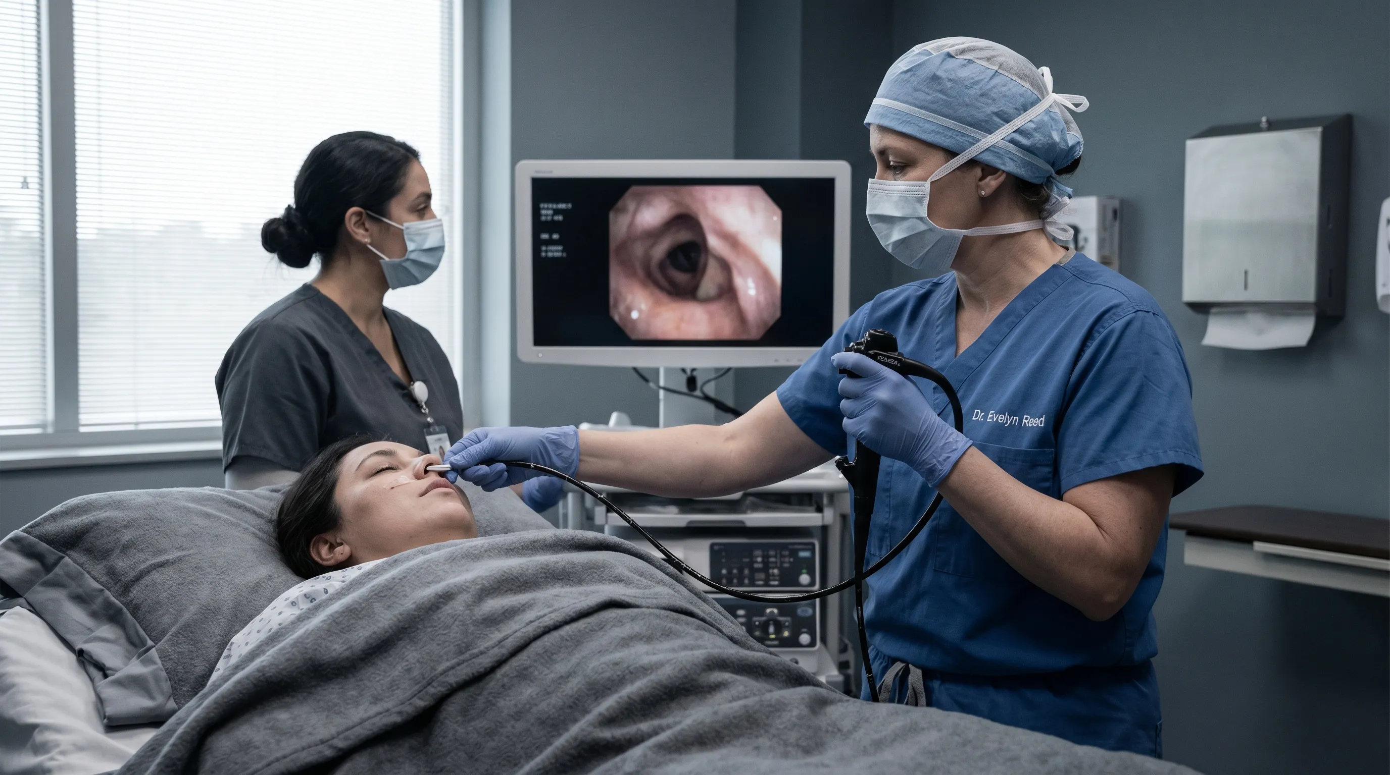
What Happens During the Bronchoscopy Procedure?
A typical bronchoscopy takes about 20 to 30 minutes, but you should plan to be at the facility for a few hours to account for preparation, monitoring, and recovery. The actual duration depends on anatomical complexity, the number of biopsies needed, and whether advanced techniques like EBUS or airway dilation are required.
- Positioning and Monitoring: You will lie comfortably on a procedure table, usually in a supine position with the head slightly elevated. Monitors will track your heart rate, blood pressure, respiratory rate, and oxygen saturation (pulse oximetry) continuously. Supplemental oxygen is administered via nasal cannula throughout the procedure.
- Applying Local Anesthesia & Sedation: The medical team will spray numbing medication to the back of your throat and inside your nostrils. Once sedated, you will feel relaxed and drowsy. A bite block may be placed in your mouth to protect the bronchoscope and your teeth.
- Inserting the Bronchoscope: The doctor will gently insert the bronchoscope through your nose or mouth. If entering via the nose, a vasoconstrictor spray may have been applied beforehand to reduce mucosal swelling and bleeding. You will be encouraged to take slow, deep breaths to help the scope pass the vocal cords smoothly.
- Visual Inspection: The doctor will navigate the scope through your trachea and into the branching bronchi, examining the airway lining on a video monitor. The mucosal surface, vascular patterns, cartilaginous rings, and bifurcations are carefully evaluated for inflammation, polyps, tumors, stenosis, or dynamic airway collapse during breathing.
- Collecting Samples: If needed, the doctor may perform a Bronchoalveolar Lavage (BAL), brushings, washings, or a biopsy. Forceps can gently pinch tiny tissue fragments, while a cytology brush collects surface cells. Samples are immediately placed in preservative solutions or sterile containers and sent to pathology and microbiology labs for analysis.
- Completion: Once the examination is complete, the doctor gently withdraws the bronchoscope. The bite block is removed, and you will be assisted to a recovery position. Suction may be used one final time to clear residual secretions.
Video: An educational video explaining the bronchoscopy examination process and what patients should expect.
Throughout the procedure, the nursing team remains at your side, monitoring your comfort, providing suction if you cough excessively, and adjusting sedation as needed. The bronchoscope does not block your ability to breathe; air flows freely around the thin instrument, and your respiratory center automatically compensates. Communication is maintained, though speaking is limited due to the scope's position.
Does a Bronchoscopy Hurt?
Fiberoptic bronchoscopy is not typically painful. The combination of local anesthetic and conscious sedation ensures most patients experience minimal discomfort. The procedure is designed to prioritize patient safety and comfort, and modern sedation protocols have significantly improved the tolerability of airway endoscopy.
- You may feel pressure as the scope is inserted, similar to swallowing a thick pill or experiencing mild throat tightness.
- You might have the urge to cough, but this is greatly reduced by the numbing medication and the sedative's antitussive effects. If coughing becomes pronounced, the physician can administer additional topical lidocaine through the working channel.
- Most patients feel very drowsy and relaxed. Many don't remember the procedure afterward due to the amnestic properties of midazolam. Some describe it as a brief nap with vague sensations of pressure.
- Afterward, you may have a mild sore throat or hoarseness for a day or two, similar to a mild upper respiratory infection. This resolves spontaneously with hydration, throat lozenges, and rest.
It is important to note that anxiety often exacerbates the perception of discomfort. Controlled breathing techniques, listening to calming music through provided headphones (if permitted), and maintaining open communication with the procedural team about your comfort level can significantly enhance the experience. Rarely, patients with severe airway hypersensitivity or complex anatomical variations may require deeper sedation or general anesthesia with endotracheal intubation to ensure safety and procedural success.
Risks and Complications of Bronchoscopy
Fiberoptic bronchoscopy is a very safe procedure with a well-documented safety profile, but like any medical intervention, it carries some small risks. The overall complication rate is less than 5%, and serious adverse events are exceedingly rare, occurring in less than 1% of cases when performed by experienced operators.
Common, Minor Side Effects:
- Sore throat or hoarseness, typically resolving within 24-48 hours.
- Coughing up small streaks of blood, especially after biopsy or BAL. This usually clears quickly.
- A low-grade fever for up to 24 hours, often a transient inflammatory response to airway manipulation or saline lavage.
- Nasal irritation or minor epistaxis (nosebleed) if the transnasal approach is used.
Less Common but More Significant Risks:
- Bleeding: More significant bleeding can occur after a biopsy, particularly in patients with coagulopathy or vascular tumors. Endoscopists manage this with topical epinephrine, cold saline, balloon compression, or electrocautery. Prophylactic correction of clotting abnormalities beforehand minimizes this risk.
- Pneumothorax (Collapsed Lung): This is a small risk specifically associated with transbronchial biopsies that penetrate the visceral pleura. It occurs in approximately 0.5-2% of cases. Symptoms include sudden sharp chest pain and shortness of breath. Most small pneumothoraces resolve spontaneously, but larger ones may require needle aspiration or chest tube placement.
- Bronchospasm or Laryngospasm: Airway irritation can trigger reflex constriction, particularly in asthmatic or COPD patients. Pre-procedure bronchodilators and adequate sedation reduce this risk. Rescue nebulizers and advanced airway management techniques are always immediately available.
- Infection: There is a very low risk of developing pneumonia after the procedure, especially in immunocompromised individuals. Strict endoscope reprocessing guidelines, adherence to aseptic technique, and prophylactic antibiotics when indicated mitigate transmission risks.
- Cardiopulmonary Complications: Transient arrhythmias, hypoxia, or blood pressure fluctuations may occur, particularly in patients with severe baseline cardiopulmonary disease. Continuous monitoring and experienced anesthesia management ensure rapid recognition and intervention.
Patients are carefully screened beforehand to identify high-risk factors, and procedural modifications are made accordingly. The benefit of obtaining an accurate diagnosis almost always outweighs the small procedural risk.
Recovery and Aftercare
Recovery is usually quick and straightforward, with most patients returning to baseline within 24 hours. Understanding the post-procedure timeline helps patients set realistic expectations and recognize normal healing versus warning signs.
- Recovery Area: You will rest for 1-2 hours in a post-anesthesia care unit (PACU) while the sedation wears off. Nurses will monitor your vital signs, assess your gag reflex, and check for immediate complications. You will receive supplemental oxygen until your baseline saturation is stable.
- Eating and Drinking: Do not eat or drink anything until the numbness in your throat is gone (usually 1-2 hours) to prevent choking or aspiration. Start with cool, clear liquids, then progress to soft foods. Avoid hot, spicy, or crunchy foods on the first day, as they can irritate the healing airway mucosa.
- Going Home: You must have someone drive you home. Avoid driving, operating heavy machinery, signing legal documents, or making important decisions for 24 hours. The sedative medications linger in your system, impairing coordination and cognitive function even if you feel fully awake.
- Activity & Lifestyle: Rest for the remainder of the day. Avoid strenuous exercise, heavy lifting, or intense cardio for 24-48 hours to minimize bleeding risk and allow airway inflammation to subside. Light walking is encouraged to promote circulation and prevent respiratory stasis.
- Symptom Management: Over-the-counter acetaminophen can be used for mild throat discomfort or headache. Avoid NSAIDs like ibuprofen for the first 48 hours if biopsies were performed, as they can interfere with platelet function. Warm saltwater gargles, humidified air, and throat lozenges provide soothing relief.
- Follow-Up: You will have a follow-up appointment to discuss biopsy results. Pathology typically takes 3-7 business days, while microbiological cultures may require 7-14 days. Your pulmonologist will integrate these findings with your clinical picture and imaging to formulate a comprehensive management plan.
When to Call Your Doctor: Contact your doctor immediately or seek emergency medical care if you experience severe chest pain, significant or worsening shortness of breath, coughing up more than a few tablespoons of bright red blood, difficulty swallowing, high fever (over 101.5°F / 38.6°C) that persists beyond 48 hours, or signs of infection such as chills, rigors, or purulent sputum production. Prompt reporting of these symptoms ensures rapid intervention and optimal outcomes.
Benefits of Fiberoptic Bronchoscopy
This procedure is valuable for several reasons, offering a unique blend of diagnostic precision and therapeutic capability that non-invasive alternatives cannot match:
- Minimally Invasive: Direct visualization of the lungs without surgical incisions or prolonged hospitalization. Compared to video-assisted thoracoscopic surgery (VATS) or open thoracotomy, bronchoscopy involves shorter procedure times, faster recovery, lower pain scores, and reduced healthcare costs.
- Accurate Diagnosis: Precise diagnosis of cancer, infection, and other diseases. Histopathological confirmation is essential for oncologic treatment pathways, antibiotic stewardship, and targeted immunosuppressive therapy. Bronchoscopy provides tissue architecture that imaging alone cannot deliver.
- Therapeutic Capabilities: Can be used to remove foreign objects, clear mucus plugs, dilate strictures, place stents, ablate tumors, and manage life-threatening hemoptysis. This dual diagnostic-therapeutic role often eliminates the need for multiple procedures or emergency surgeries.
- Advanced Technology Integration: Techniques like EBUS (Endobronchial Ultrasound), navigational bronchoscopy with electromagnetic tracking, robotic-assisted platforms, and rapid on-site evaluation (ROSE) by cytopathologists have dramatically expanded its diagnostic reach, particularly for peripheral lung nodules that were previously inaccessible.
- Cost-Effectiveness & Accessibility: Performed in outpatient settings, it reduces inpatient admissions, shortens diagnostic timelines, and accelerates the initiation of appropriate treatment. It is widely available in regional medical centers and covered by most insurance plans when medically indicated.
Conclusion
Fiberoptic bronchoscopy is a safe, highly effective, and indispensable tool for diagnosing and treating a wide spectrum of pulmonary conditions. While the thought of having a scope passed through your airway may initially seem daunting, decades of clinical refinement, advanced sedation protocols, and meticulous procedural standards have made it a routine, well-tolerated intervention with minimal discomfort for the vast majority of patients. Understanding the clinical indications, preparation steps, procedural sequence, and aftercare guidelines empowers you to actively participate in your respiratory healthcare journey.
By providing clear visual insights and reliable tissue samples, bronchoscopy often marks a critical turning point toward an accurate diagnosis, targeted treatment, and improved quality of life. Open communication with your pulmonology team, strict adherence to pre- and post-procedure instructions, and timely reporting of symptoms ensure the safest possible outcome. Ultimately, this procedure bridges the gap between suspicion and certainty, equipping you and your physicians with the information needed to navigate your lung health with confidence and precision.
Frequently Asked Questions
How long does it take to get bronchoscopy results back?
The timeline for results depends on the type of samples collected and the specific laboratory tests ordered. Preliminary visual findings are discussed immediately after the procedure. Rapid on-site cytology (ROSE), if used, can provide preliminary cancer or infection screening within 15-30 minutes. Standard tissue pathology reports typically take 3 to 7 business days to finalize, as they require tissue processing, staining, and expert review. Microbiological cultures for bacteria, fungi, or mycobacteria (like TB) require longer incubation periods and may take 7 to 21 days. Your care team will contact you as soon as definitive results are available and schedule a follow-up visit to discuss next steps.
Is general anesthesia required for a bronchoscopy, or is conscious sedation enough?
In the majority of cases, conscious sedation combined with topical airway anesthesia is sufficient and preferred. Conscious sedation allows you to remain awake enough to follow simple instructions (like taking deep breaths) while keeping you comfortable, amnesic, and relaxed. General anesthesia with endotracheal intubation or a laryngeal mask airway is reserved for specific scenarios, such as prolonged therapeutic procedures (e.g., complex stent placement or airway tumor debulking), patients with severe respiratory compromise who cannot tolerate spontaneous breathing during the procedure, or individuals with extreme anxiety or anatomical challenges. Your pulmonologist and anesthesia provider will tailor the plan to your clinical needs.
What should I do if I cough up blood after the procedure?
Coughing up small streaks of blood or rust-tinged mucus for up to 48 hours after bronchoscopy is common, especially if a biopsy was performed. This occurs because the airway mucosa is highly vascular and the biopsy forceps create tiny, controlled wounds that ooze as they heal. Stay hydrated, rest, and avoid vigorous coughing, heavy lifting, or blood-thinning medications unless prescribed. Use a humidifier and sip cool fluids to soothe the airway. However, if you cough up more than a few tablespoons of bright red blood, notice clots, experience worsening shortness of breath, dizziness, or a rapid heart rate, seek immediate medical attention, as these may indicate significant hemorrhage requiring clinical intervention.
Are there alternatives to bronchoscopy for diagnosing lung nodules or infections?
Yes, but they often have different risk-benefit profiles and diagnostic yields. CT-guided transthoracic needle biopsy is an alternative for peripheral lung lesions, though it carries a higher risk of pneumothorax (collapsed lung) and cannot visualize the central airways. Sputum cytology and culture are non-invasive but have low sensitivity, particularly for early-stage cancers or localized infections. Video-assisted thoracoscopic surgery (VATS) or surgical lung biopsy provides larger tissue samples and is considered the gold standard for diffuse interstitial lung diseases, but it requires general anesthesia, longer hospitalization, and carries higher morbidity. Bronchoscopy often serves as the optimal first-line invasive test due to its versatility, safety, and ability to combine sampling with direct visualization.
Can I drive myself home after a fiberoptic bronchoscopy?
No, you cannot drive yourself home or for at least 24 hours after the procedure. The intravenous sedatives used (typically benzodiazepines and opioids) significantly impair your reaction time, coordination, judgment, and visual processing, even if you feel completely awake and alert. These effects can persist longer than the subjective feeling of drowsiness. Additionally, local anesthetic in your throat can affect your gag reflex temporarily, and your overall stamina may be reduced. You must arrange for a responsible adult to accompany you to the appointment, stay in the waiting area, and drive you home. Failure to follow this guideline not only poses serious safety risks to yourself and others but may also violate medical discharge protocols and insurance requirements.
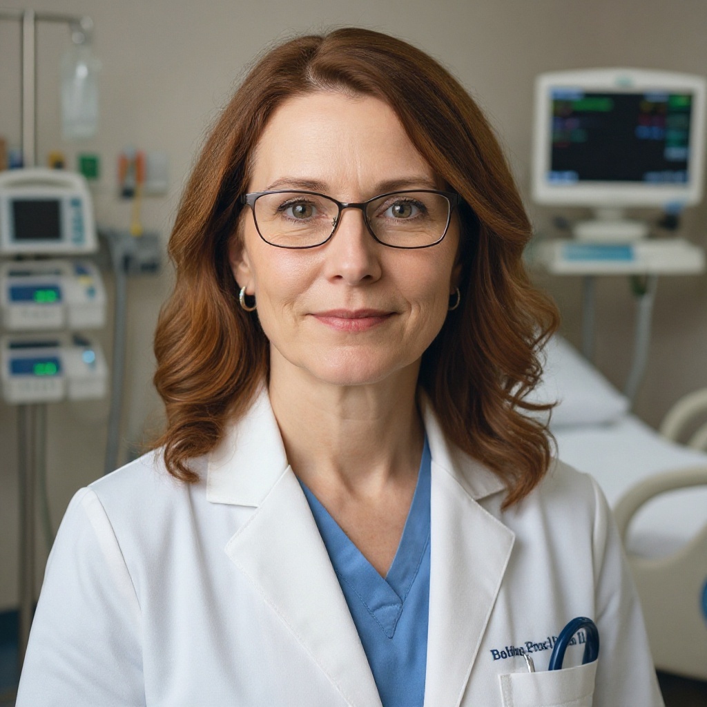
About the author
Evelyn Reed, MD, is double board-certified in pulmonary disease and critical care medicine. She is the Medical Director of the Medical Intensive Care Unit (MICU) at a major hospital in Denver, Colorado, with research interests in ARDS and sepsis.